Our Bureau
Newark, NJ
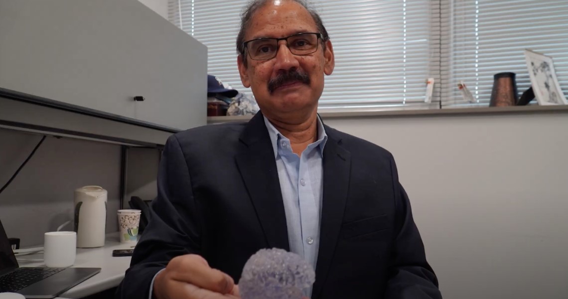
New Jersey Institute of Technology, NJIT has announced the name of Bharat Biswal as this year’s winner of NJIT’s Excellence in Research award. Bharat Biswal, a pioneer in the field of neural imaging who developed a technique that sheds new light on brain-related diseases and injuries.
Biswal thanked the selection committee and senior administration at NJIT for choosing him for the award. He added, “But I’m particularly grateful to all the undergraduate researchers who, in spite of their very busy schedules, still make time to do research and contribute very meaningful work, and to the graduate students, postdocs and colleagues who helped me in this research.”
Biswal joined NJIT in 2012 as a tenured full professor and was promoted to distinguished professor in 2016. That same year, he was elected a fellow of the American Institute of Medical and Biological Engineering. He has received continuous extramural funding since joining NJIT of more than $8 million and has more than 48,000 citations, while his most-referenced paper received 10,548 citations.
Atam Dhawan, NJIT’s senior vice provost for research, said Biswal “exemplifies the qualities of a leader as a visionary in neuroscience.” Biswal’s research in the field of brain imaging have catalyzed a paradigm shift in the understanding of the resting human brain and its networks, fundamentally altering how scientists and clinicians approach the study and treatment of neurological and psychiatric conditions.
In the early 1990s, “It was perceived as a crazy idea in neuroscience, which was focused on task and response,” recalled Biswal, then a graduate student at Wisconsin Medical Center and now a distinguished professor of biomedical engineering. “As an engineer, I wanted to understand the entire system of neural networks — and their connections — at baseline conditions. With one scan, we could observe activity in the motor, visual and speech regions, for example.”
Thirty years later, what is known as resting-state brain connectivity is the primary vehicle for examining brain connections in infants, very young, elderly and neurologically impaired people who can’t perform tasks on command. It is one of three required approaches in the National Institutes of Health’s Human Connectome Project.
Biswal, director of NJIT’s Center for Brain Imaging, is now using it to compare neural signaling in people with Alzheimer’s disease with those aging normally. He is zeroing in on the connections between the gray matter of the brain and the white matter. The study is among the first to perform a comprehensive analysis of white matter function using fMRI and its effects on cognition in the two groups.
As part of his study, he’s collaborating with groups around the world to gather a vast and diverse data set of more than 100,000 subjects who’ve undergone fMRI scans. He’s observing the tests to ensure that testing methodologies and practices are as similar as possible. His team is working on a set of toolkits to track and explain the progression of Alzheimer’s to give clinicians a window into who might be at risk, for example, and to assess what therapies might be effective at different stages of the disease.


